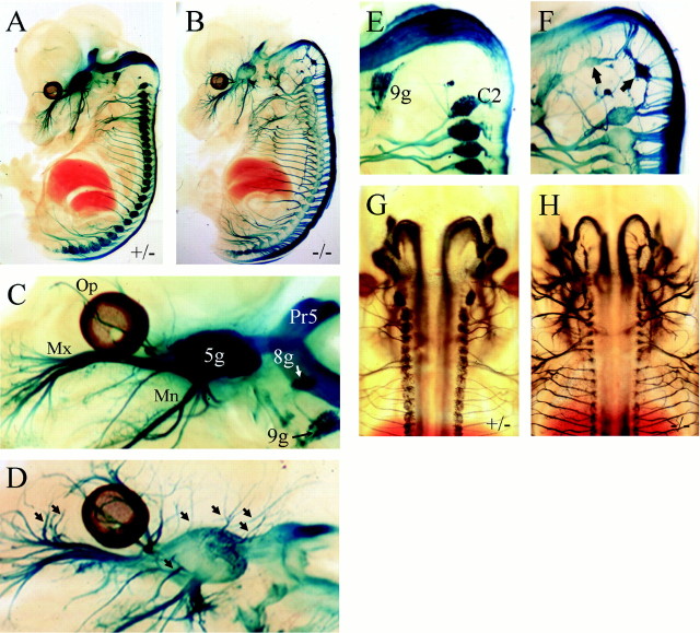
Fig. 3.
Sensory axon growth in Brn3a null mice. Brn3a+/− control (A,C, E, G) and Brn3a−/− (B, D,F, H) E13.5 embryos were stained for β-gal activity and (A–F) were hemisected before clearing. The major axonal projections of the sensory ganglia are present in Brn3a knock-out mice (A,B), but there is extensive abnormal branching, particularly in the ophthalmic and maxillary divisions of the trigeminal (C, D, arrows). Occipital sensory structures, which normally regress by this stage (E), persist abnormally in Brn3a null mice (F, arrows). Dorsal views of control and Brn3a knock-out embryos show that the aberrant sensory axons do not cross the midline in either the CNS or the periphery (G,H). The patterns of axonal growth in 11 kb/LacZ, Brn3a−/− and heterozygous controls were very similar in specimens obtained from three different litters staged from E13.5 to E14.5. In preliminary experiments, it was observed that Brn3a−/− embryos stained more intensely than Brn3a+/− littermates, consistent with negative autoregulation by Brn3a. Because we wished to ensure that no axonal projections would be overlooked in control embryos as a result of lower reporter expression, controls were deliberately stained two to four times longer than knock-out embryos. This overstaining resulted in very intense signal in the sensory ganglia of controls (A, C) and some diffusion of the β-gal reaction product into the surrounding tissue of the ganglia.5g, Trigeminal ganglion; 8g, vestibulocochlear ganglion; 9g, superior ganglion of glossopharyngeal nerve; C2, dorsal root ganglion corresponding to cervical segment C2; Mn, mandibular division of trigeminal nerve; Mx, maxillary division of trigeminal nerve; Op, ophthalmic division of trigeminal nerve; Pr5, principal trigeminal nucleus.

