
Fig. 7.
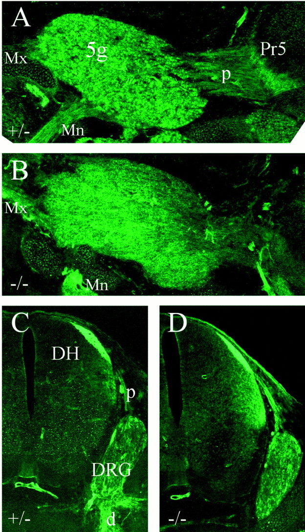
Neuropilin-1 expression in control and Brn3a-deficient mice. Brn3a+/− (A,C) and Brn3a−/− (B,D) embryos were examined at E14 for Npn1 immunoreactivity. Sagittal sections through the trigeminal ganglion and hindbrain (A, B) and cross-sections through the cervical spinal cord and dorsal root ganglion (C, D) show Npn1 expression in the expected sensory structures in both the presence and absence of Brn3a.5g, Trigeminal ganglion; d, distal root (of ganglion); DH, dorsal horn (of spinal cord);DRG, dorsal root ganglion; Mn, mandibular division, trigeminal nerve; Mx, maxillary division, trigeminal nerve; p, proximal root (of ganglion).