
Fig. 2.
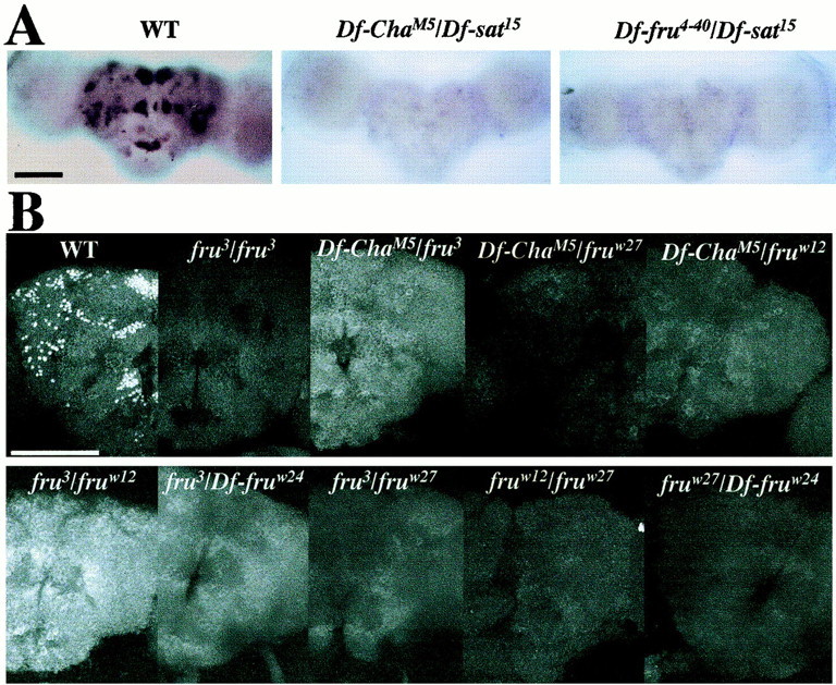
Lack of sex-specific fruitlessexpression in the CNS of fru-breakpoint mutants. A, In situ hybridizations performed on pupal progeny resulting from crosses involving three of the deletions depicted in Figure 1; 1-d-old male pupae had afru-derived riboprobe (Fig. 1) applied to whole-mounted CNSs of wild type (WT, n = 11) and these twoDf/Df types (Df-ChaM5/Df-sat15,n = 6;Df-fru4–40/Df-sat15,n = 6); no signals were elicited by this nucleic-acid probe in any of the 12 double-deletion specimens.B, Anti-FRUM immunohistochemistry performed on pupal progeny resulting from crosses of various deletions and other breakpoint variants (Fig. 1); heterozygotes involving certain of the chromosome aberrations and one of the frutransposon mutants were included as a negative control (compare Fig. 3; Table 2); antibody against the male-specific form of the protein was applied to whole-mounted CNSs dissected from 2-d-old male pupae. Summaries of these immuno-histochemical results (including numbers of samples per genotype) are given in Table 1. Signals (or the absence thereof) were examined by confocal microscopy, and representative images were made for specimens of the various genotypes at 40×.A, B, anterior views of the brains.B, The brain image offru3/fruw12(lower left panel) shows a whitishgeneral background staining that was not detected in other specimens of this genotype (cf. Table 1) and bears no relation to the WT pattern. Scale bars: A, 100 μm; B, 50 μm.