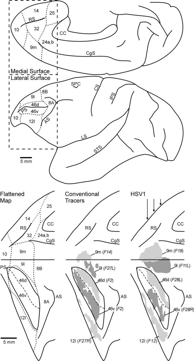
Fig. 1.

Location of HSV1 and conventional tracer injections. Top, The lateral surface and medial wall of the cerebral cortex of a cebus monkey. Bottom, The lateral surface and medial wall of the frontal lobe (boxed-in area above) aligned on the midline between the two and both banks of the principal sulcus unfolded.Left, A flattened map of the cytoarchitectonic regions of the prefrontal cortex according to the criteria of Walker (1940) andBarbas and Pandya (1989). Dashed lines indicate the approximate location of the borders between these regions.Middle, The reconstructed injection sites for different conventional tracer experiments. Right, Selected HSV1 injection sites. Shading is used to indicate the combined zones I and II of each injection site, and any regions of overlap are indicated with dotted lines. The approximate locations of the coronal sections through the HSV1 injection sites (see Fig. 2) are indicated with vertical arrowsin the right panel. AS, Arcuate sulcus;CC, corpus callosum; CS, central sulcus;CgS, cingulate sulcus; IPS, intraparietal sulcus; LS, lateral sulcus; PS, principal sulcus; RS, rostral sulcus; SPC, superior precentral sulcus; STS, superior temporal sulcus.
