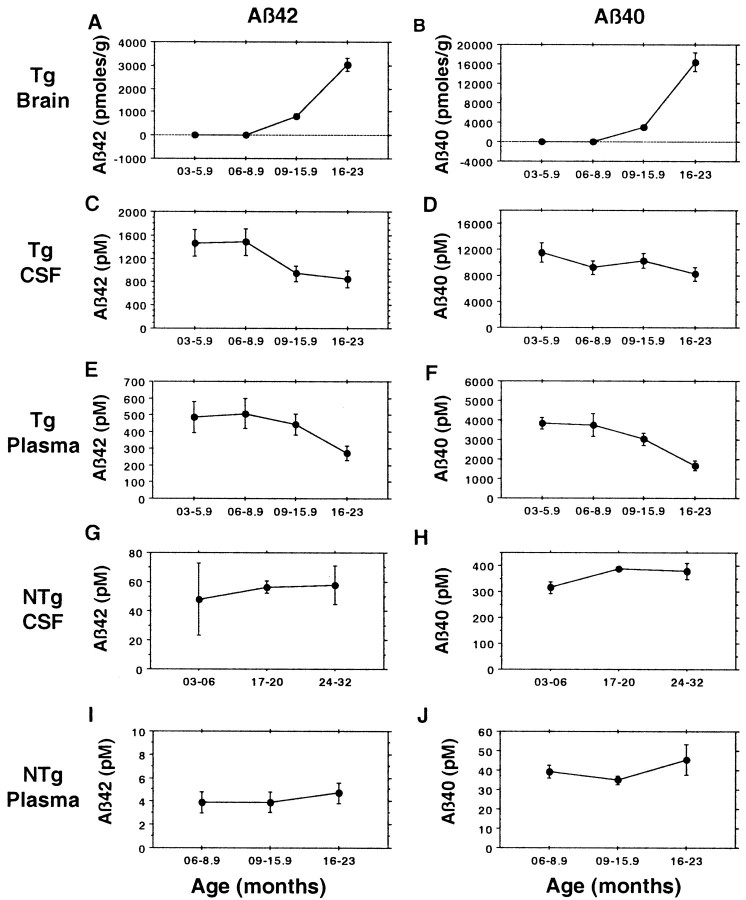
Fig. 7.
CSF and plasma Aβ in Tg2576 mice decline as Aβ is deposited in the brain. Total brain Aβ42 (A) and Aβ40 (B) were assayed by 3160/BC05 or 3160/BA27 ELISAs, respectively; see also Figure 2 and Table 2. Tg2576 CSF and plasma Aβ42 (C, E) were assayed by BNT77/BC05 ELISA, and Tg2576 CSF and plasma Aβ40 (D, F) were assayed by BAN50/BA27 ELISA. Both nontransgenic (NTg) CSF and plasma Aβ42 (G, I) and Aβ40 (H, J) were assayed with BNT77 capture. The number of Tg2576 CSF samples assayed for the four time groups are 9, 9, 16, and 11, totaling 45. The number of Tg2576 plasma samples assayed for the four time groups are 30, 18, 64, and 19, totaling 131. The decline for CSF Aβ42 is significant (p = 0.02), and the declines for plasma Aβ40 and Aβ42 are highly significant (Aβ42, p= 0.008; Aβ40, p = 0.006; Spearman's rank correlation for the 6–23 month age range). The number of nontransgenic CSF samples assayed for the three time groups are 2, 2, and 6, totaling 10. The number of nontransgenic plasma samples assayed for the four time groups are 7, 6, 12, and 6, totaling 31.

