Fig. 5.

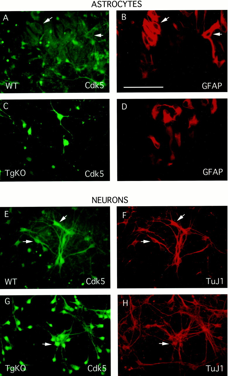
Mixed neuronal–astrocytic cultures were prepared from E17.5 WT and TgKO mice embryos as described by Vicario-Abejon et al. (1998), with minor modifications. After fixation with 4% PFA, double immunocytochemistry was performed on the cultures with either GFAP (an astrocytic marker) and Cdk5 or TuJ1 (a neuronal marker) and Cdk5. Panels on the left (FITC,green) show the staining pattern of Cdk5 with the corresponding GFAP or TuJ1 (rhodamine, red) staining to the right. WT mice show Cdk5 staining in neurons as well as astrocytes (A, B,E, F), whereas there is no Cdk5 seen in the astrocytes of TgKO mice (C,D, G, H). In addition, the levels of transgenic Cdk5 in TgKO neurons is higher than that seen in neurons derived from WT mice (compare E,G). Scale bar (in B), 100 μm.
