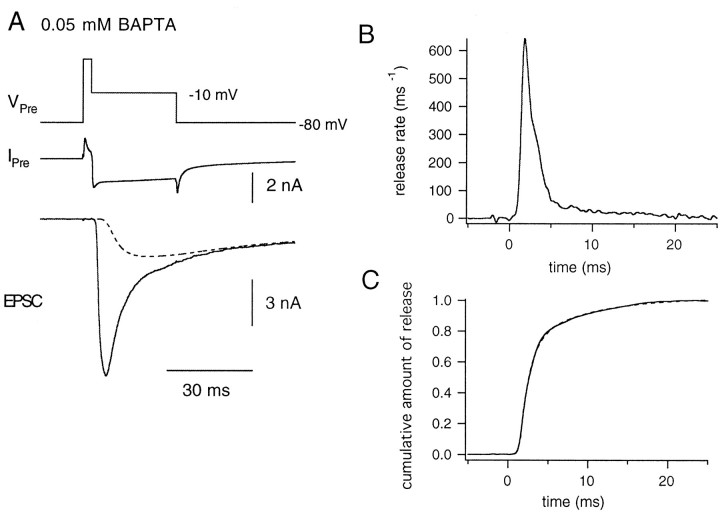
Fig. 8.
Time course of quantal release during long pulses in the presence of 0.05 mm BAPTA in the patch pipette.A, The presynaptic terminal was depolarized from −80 to +70 mV for 4 msec and was held at −10 mV for 20 msec (VPre) to elicit presynaptic Ca2+ current (IPre). The dotted line shows an estimate of the residual current component.B, Release rate plotted against time. Starting at the time point of 0, the presynaptic terminal was held at −10 mV.C, The cumulative fraction of vesicles released is plotted against time. The data could be fitted with double exponentials with time constants of 1.09 msec (68%) and 5.92 msec (dotted line).

