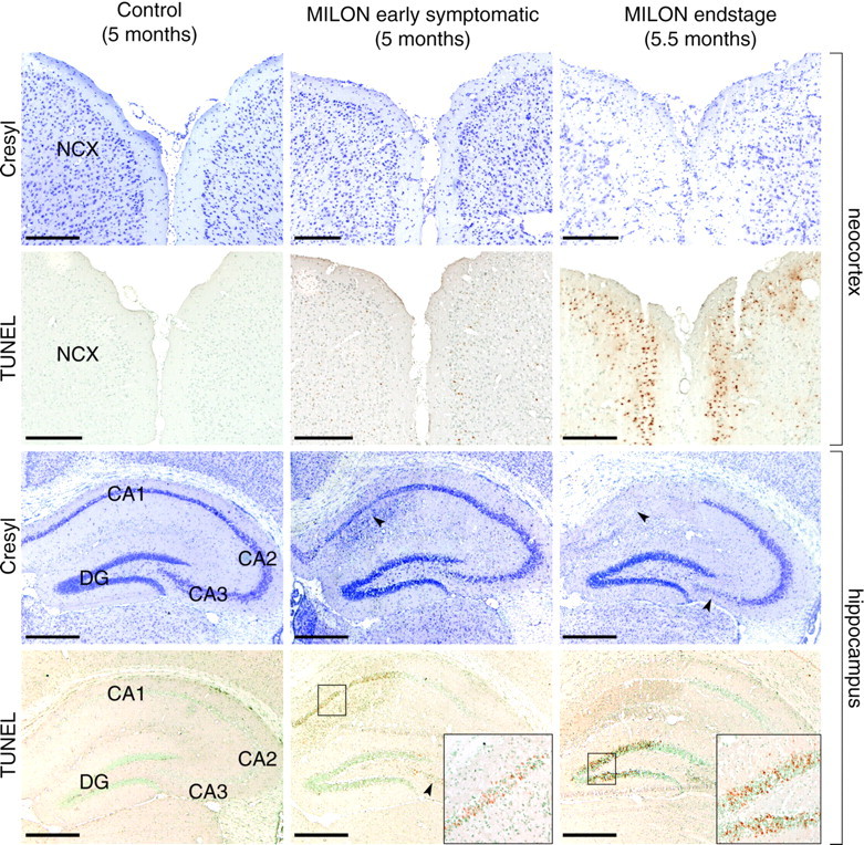
Fig. 3.

Characterization of cell loss in neocortex and hippocampus of a 5-month-old control mouse, a 5-month-old early symptomatic MILON mouse, and a 5.5-month-old end-stage MILON mouse stained with cresyl violet or TUNEL. NCX, Neocortex;DG, dentate gyrus The arrowheads indicate cell loss in the CA1 and CA3 regions. The boxes are close-ups of indicated areas. Scale bars, 0.5 mm. There is cell loss and cell infiltration in the CA1 area of hippocampus of early symptomatic MILON mice. There is extensive cell loss and degenerative changes in neocortex and in the CA1, CA3, and dentate gyrus areas of the hippocampal formation of end-stage MILON mice.
