
Fig. 4.
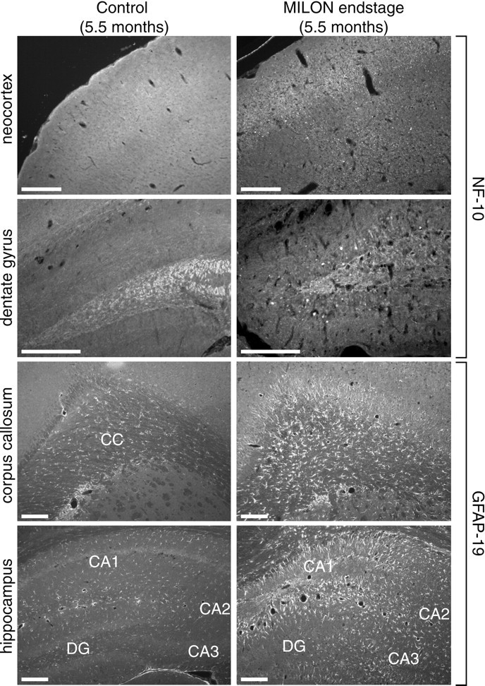
Characterization of neurodegenerative changes and gliosis in a MILON and a control mouse at the age of 5.5 months. Neurofilaments were detected with a polyclonal antibody against NF-10, and astrocytes were detected with a polyclonal antibody against GFAP. Antibody reactivity was visualized with immunofluorescence.CC, Corpus callosum; DG, dentate gyrus. Scale bars, 0.25 mm. NF-10 immunofluorescence shows extensive axonal beading in neocortex and accumulation of neurofilaments in granular cell somata of dentate gyrus in MILON mice. GFAP immunofluorescence shows extensive gliosis in corpus callosum and hippocampus.