
Fig. 3.
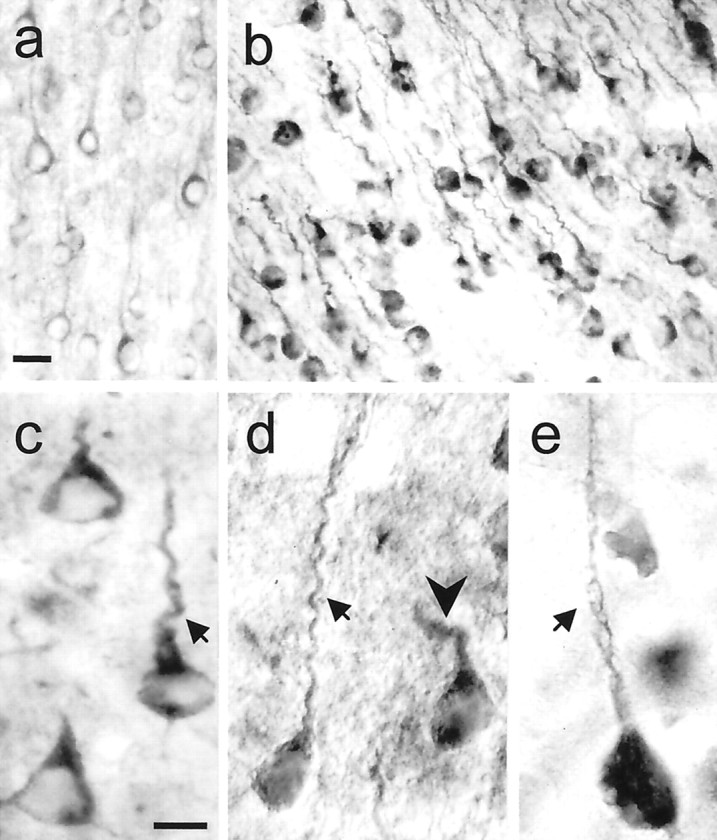
Increased huntingtin immunoreactivity and dysmorphic dendrites in HD mice. a, WT mouse (8.5 months, rotarod, normal behavior) exhibits low to moderate levels of huntingtin labeling in the cytoplasm and proximal dendrites of pyramidal neurons. b–e, HD mice have intense labeling for huntingtin in the cytoplasm and proximal dendrites (b, HD100, L63, 4.5 months, RR = 10, +clasp;c, HD46, L17, 12 months, RR = 66, +clasp;d, HD100, L63, 7 months, RR = 51; e, HD100, L46, 12 months, RR = 26). Some neurons also show diffuse nuclear staining (d). Dysmorphic dendrites show marked retraction (arrows in c, d) and disorientation (arrowhead in d). Membrane blebbing occurs in cell bodies and dendrites (arrow ine). Staining was performed with anti-huntingtin antibody Ab585. Scale bars: a, b, 25 μm;c–e, 10 μm.