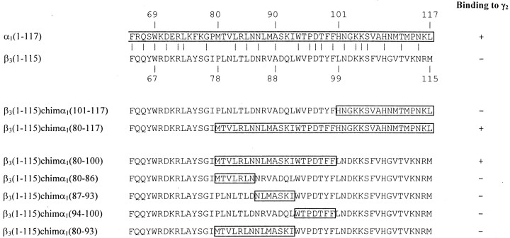
Fig. 2.
α1(80–100) forms the contact site to γ2 subunits. C-terminal sequences of the fragments α1(1–117), β3(1–115), and of different chimeras are shown. Amino acid sequences of the α1subunit are boxed. HEK cells were cotransfected with these constructs together with γ2 subunits, and a possible coimmunoprecipitation was investigated, as described in Results. + indicates binding, and − indicates absence of binding between these constructs and full-length γ2subunits. The experiments were performed three times with similar results.

