Fig. 3.
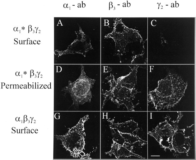
Immunofluorescence of HEK cells cotransfected with GABAA receptor subunits. HEK cells were cotransfected with α1*, β3, and γ2subunits (A–F) or with α1, β3, and γ2 subunits(G–I). Immunofluorescence was performed using α1(1–9) antibodies (A, D, G), β3(1–13) antibodies (B, E, H), or γ2(1–33) antibodies (C, F, I) on the cell surface (A–C, G–I) or in permeabilized cells(D–F) by confocal laser microscopy (single sections). Scale bar, 10 μm. The experiment was performed four times with similar results.

