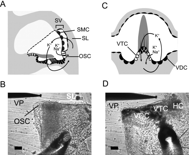
Fig. 1.
Tissue preparation for OSC and VTC.A, C, Schematic illustration showing the location of OSC and VTC in the cochlea and semicircular canal ampulla, respectively.B, D, Prepared tissue for the measurement ofIsc in OSC and VTC, respectively.HC, Vestibular hair cell (damaged); SL, spiral ligament; SMC, strial marginal cell;SV, stria vascularis; VP, vibrating probe. Scale bar, 50 μm. A andC adapted from graphics by P. Wangemann (Wangemann, 1997).

