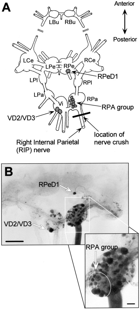
Fig. 1.

Experimental model and nerve injury procedure.A, Schematic representation of the dorsal aspect of theLymnaea CNS with the cerebral commissure cut and cerebral ganglia folded outward. LBu/RBu, Left and right buccal ganglia; LCe/RCe, left and right cerebral ganglia; LPe/RPe, left and right pedal ganglia;LPl/RPl, left and right pleural ganglia;LPa/RPa, left and right parietal ganglia;Vi, visceral ganglion; RPeD1, right pedal dorsal 1; RPA, right parietal A neurons;RPD1, right parietal dorsal 1; VD2/VD3, visceral dorsal 2 and 3 [nomenclature according to Benjamin and Winlow (1981)]. B, Microphotograph of aLymnaea CNS after retrograde staining of the RIP nerve (buccal ganglia not shown). Scale bar, 500 μm. In this case no nerves were crushed. Backfilling the RIP nerve labeled numerous neuronal somata. Particularly, RPeD1, VD2/VD3, and the RPA group motoneurons are clearly visible (also see enlarged inset of RPa ganglion). Scale bar, 100 μm.
