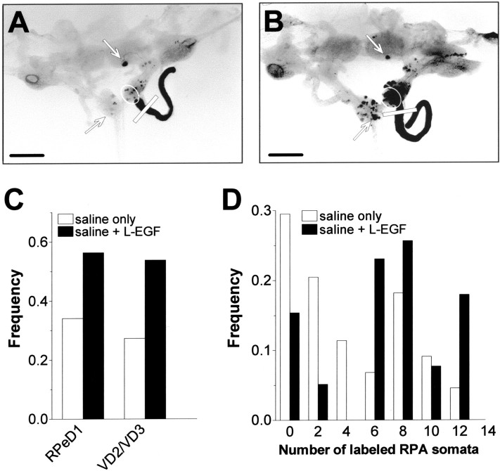
Fig. 5.
Effect of L-EGF on regeneration of crushed RIP axons. A, B, Microphotographs of isolated CNSs (dorsal view, with the cerebral commissures cut) that were cultured for 2 d after receiving a crush to the RIP nerve in saline (A) or in saline plus 100 nmL-EGF (B). Scale bar, 500 μm. Comparison of both photographs illustrates that the number of labeled neuronal somata was dramatically enhanced in the presence of L-EGF. C, Frequency of preparations in which RPeD1 and VD2/VD3 were retrograde labeled after 2 d in the presence of 100 nm L-EGF (saline + L-EGF) and without (saline only). Treatment with L-EGF significantly enhanced the proportion of preparations that extended axons from RPeD1 and VD2/VD3 into the damaged RIP nerve. D, Frequency distribution of labeled RPA somata per CNS in preparations that were cultured for 2 d in the absence (saline only) and presence of 100 nm L-EGF (saline + L-EGF) (bin width = 2). Treatment with L-EGF significantly enhanced regeneration of RPA axons projecting into the RIP nerve.

