
Fig. 5.
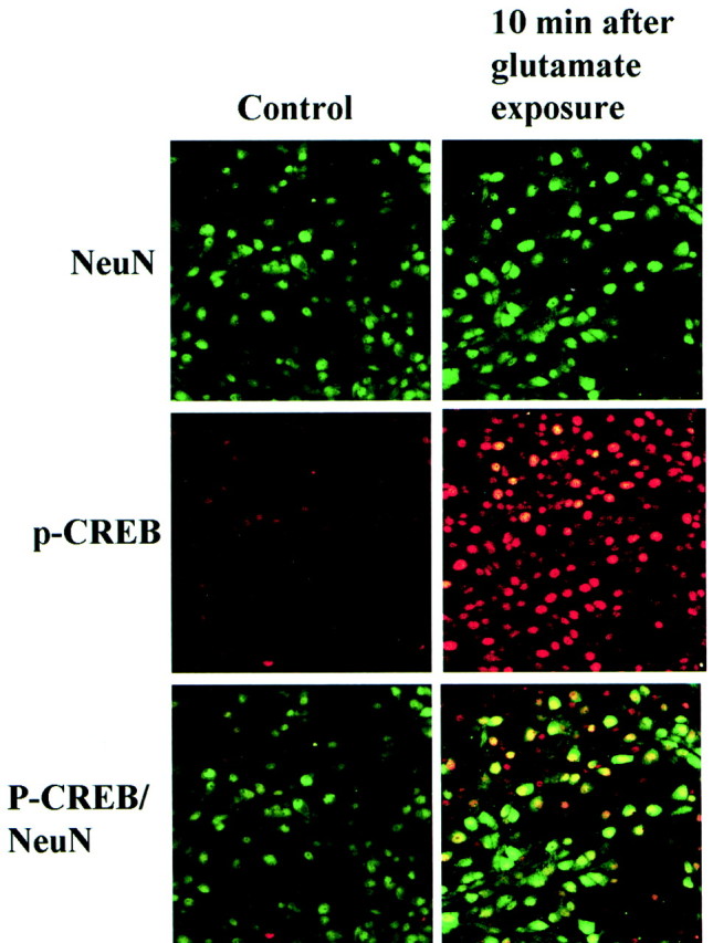
pCREB in cultured neurons after exposure to glutamate. NeuN (green) and pCREB (red) immunofluorescence in cultured hippocampal neurons 10 min after exposure to glutamate (right column) compared with controls (left column).Top, The number of neurons stained with an anti-NeuN antibody (green) was unchanged.Middle, The number of pCREB-positive cells (red) increased markedly 10 min after exposure to glutamate. Bottom, NeuN-positive–pCREB-positive cells (yellow). CREB phosphorylation was enhanced in most neurons after exposure to glutamate.