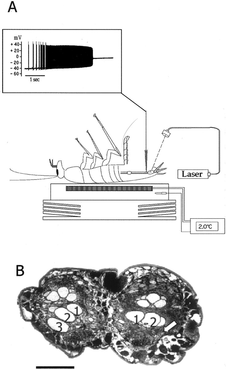
Fig. 1.

The photoablation procedure. A, Drawing of a cockroach placed ventral side up and anesthetized by cooling on a Peltier device. A cuticle flap was opened to access the ventral nerve cord for intracellular recording and ionophoresis of carboxyfluorescein. Inset, Laser illumination induced a tonic burst of action potentials and subsequent loss of membrane potential [modified from Libersat and Mizrahi (1996)].B, A paraffin section of the ventral nerve cord of a GI3-ablated animal showing the axonal profiles of the GIs (numbers indicate the axons of GI1, GI2, and GI3, respectively); thearrow indicates the position of the missing axonal profile of right GI3. Scale bar, 100 μm.
