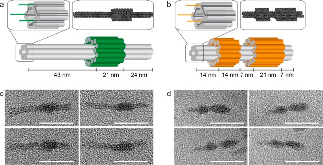
Figure 2.
DNA nanostructure design and characterization. (a,b) Representations of DNA nanostructures A (a) and B (b). The core of each structure is a 6-helix bundle (each double helix is represented by a gray cylinder). Staple extensions provide three single-stranded handle sequences at one end of the six-helix bundle (top left insets). The valency can be controlled by replacing one or more handles with a dummy sequence unrelated to that of the peptide-functionalized oligonucleotide tags. Asymmetrically positioned sleeves wrapped around the 6-helix bundle (green in (a) and orange in (b)) allow the two structures to be distinguished by TEM. Top right insets: atomic models of origamis A (a) and B (b) predicted by CanDo and visualized with QuteMol. (c,d) TEM images confirming the formation of origamis A (c) and B (d). Scale bars: 50 nm.

