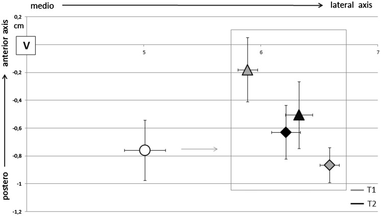
Figure 3.
CoGs position in respect with vertex for controls (circular marker) and MS subjects (triangle for the unaffected side and rhombus for the affected side) at baseline (T1) and follow-up (T2). CoGs in MS subjects were laterally displaced in comparison with controls (p < 0.0001). At baseline, CoG of the unaffected side was more medially (p < 0.0001) and anteriorly (p = 0.03) positioned than in the affected side in MS. At follow-up CoGs positions of the two hemispheres were more symmetric.

