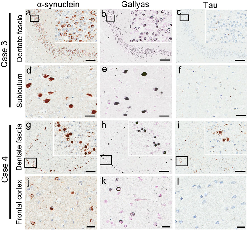
Fig. 3.
Neuronal inclusions are intensely positive on α-synuclein (a, d, g, j) and Gallyas silver stains (b, e, h, k), but tau immunoreactivity is variable among each case and region. In Case 3, almost no neuronal inclusions are positive on tau stains in the hippocampal dentate fascia (c) or subiculum (f). In Case 4, some of neuronal inclusions are positive on tau stains in the hippocampal dentate fascia (i), while no neuronal inclusions are positive on tau stains in the frontal cortex (l). Scale bars a–c, g–i; 200 μm, d–f, j–l; 50 μm

