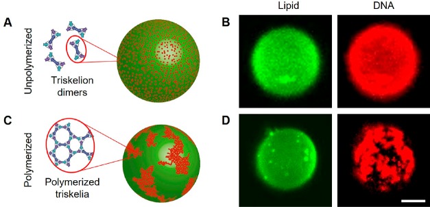
Figure 2.
DNA triskelia interacting with giant unilamellar vesicles. (A, C) Inferred distributions of DNA triskelion dimers on GUVs. Unpolymerized DNA triskelia are homogeneously distributed on the GUV surface; polymerization, triggered by addition of polymerization staples, causes triskelia to assemble into arrays in mesoscopic domains. (B, D) Confocal micrographs corresponding to a 500 nm thick section through the top of the GUV. The formation of large clusters of curved triskelia on polymerization is evident but has no significant effect on the lipid distribution. Scale bar: 10 μm.

