Figure 5.
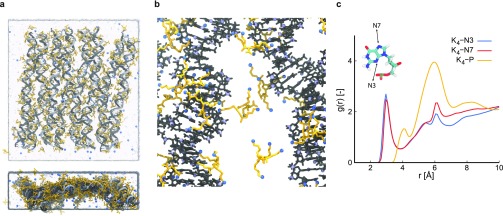
PLL-4 simulation. (a) Snapshot of the final configuration in two orientations, with the DNA origami shown in gray, water in light blue and Na+ and K4 in blue and yellow, respectively. (b) Zoomed in simulation snapshot illustrating typical binding of K4, with DNA origami shown in gray, K4 in yellow, and nitrogen in blue. (c) Radial distribution function between nitrogens in K4 and minor groove atoms, major groove atoms, and phosphorus atoms in the DNA backbone, respectively. The inset indicates the minor (N3) and major (N7) groove atoms used in the case of guanine.

