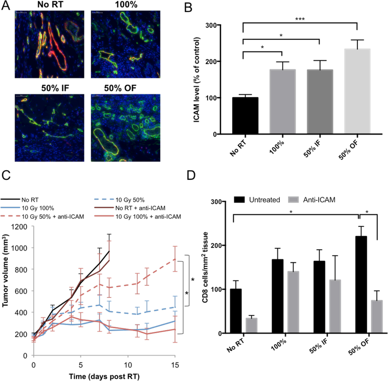
Figure 3 –
(A) Representative images of ICAM expressed on blood vessels in the tumor (red: Meca-32, green: ICAM). An increase in ICAM expression was observed 24 hours post 10Gy RT in the out-of-field half of the tumor. (B) Quantitation of ICAM staining in tumors. An average of 3 separate experiments is shown, with a total of 15 mice per group. (C) The effects of blocking ICAM on 67NR tumor response to RT. IP injections of ICAM-blocking antibody were administered at a dose of 300μg/injection 2, 16 and 48 hours after RT. Experiment was done once, with 5 mice per group. (D) Effect of blocking ICAM on CD8+ T cell infiltration following 10Gy hemi- or total irradiation and treatment with anti-ICAM antibody. IP injections of ICAM-blocking antibody were administered at a dose of 300μg/injection, 2 and 16 hours after RT. After 24 hours tumors were collected and CD8+ cells were visualized using immunofluorescence. Experiment was done once, with 5 mice per group.

