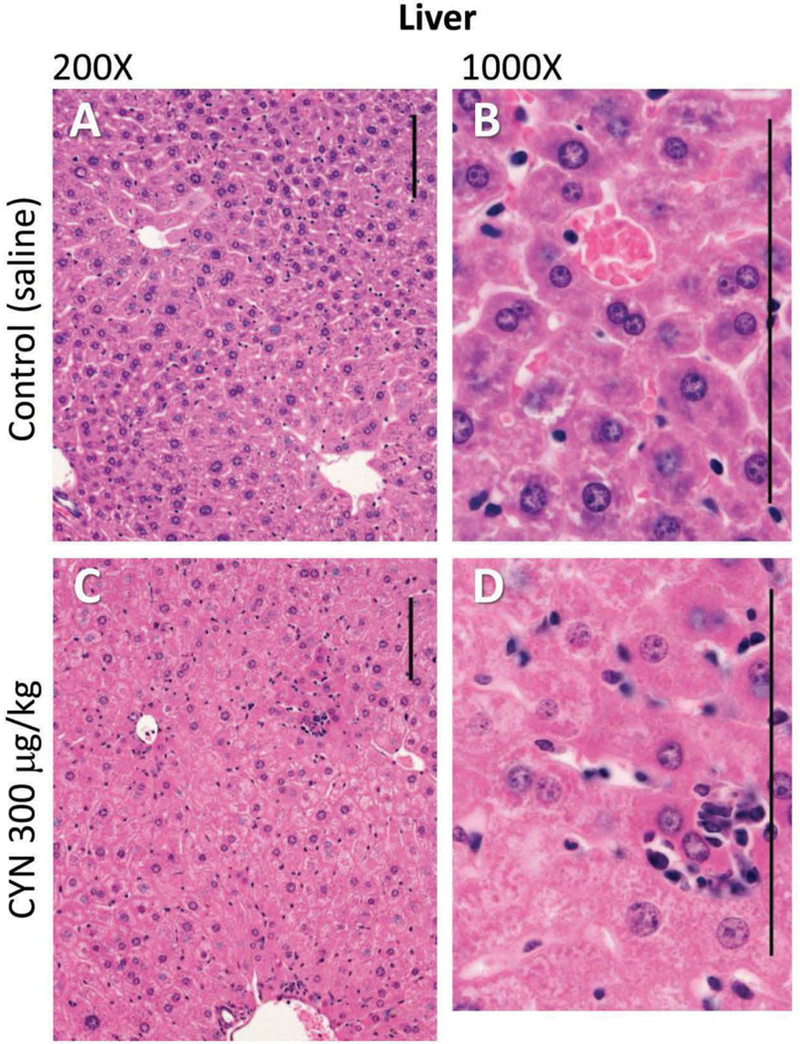
Figure 1.

Photomicrographs of the liver tissue of female mice (controls A and B; CYN 300 μg/kg C and D). Liver lesions included distortion of hepatic architecture, hepatocellular hypertrophy, cytoplasmic alteration and degeneration, and/or necrosis of centrilobular hepatocytes often infiltrated by inflammatory cells. Hematoxylin and eosin stain used. Size bars = 100 μm.
