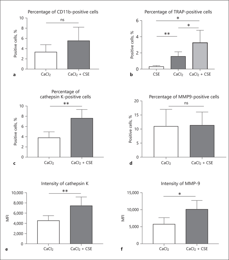
Fig. 6.

Dissociated cells from mouse aneurysmal samples collected 1 week after induction surgery were examined by flow cytometry. a To investigate myeloid cell infiltration, we used CD11b as surface marker. b To evaluate the effect of CSE, we applied CSE without CaCl2 to carotid arteries to compare TRAP-positive cells with CaCl2− and CaCl2+CSE-treated groups. In further analysis, the percentages of live cells positive for cathepsin K (c) were higher in the CaCl2+CSE group than the CaCl2 group. d On the other hand, there was no significant difference in the percentage of MMP-9-positive cells between the groups. Among the TPMs, the expression levels of cathepsin K (e) and MMP-9 (f) were higher in the CaCl2+CSE group than in the CaCl2 group. Values are presented as means ± SD for at least 3 replicates. * p < 0.05, ** p < 0.01. ns, not significant; CSE, cigarette smoke extract; TRAP, tartrate-resistant acid phosphatase; MMP-9, matrix metalloproteinase-9; MFI, median fluorescence intensity; SD, standard deviation.
