Figure 1.
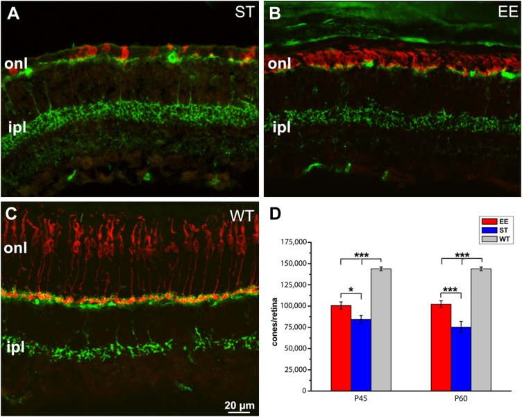
Enhanced cone survival upon exposure to enriched environment. A, B) Vertical sections of the retina of rd10 mice born and maintained in ST conditions (A) or in an enriched environment (B). C) retina of a WT control mouse. Age is 60 d (P60). Red staining: cone arrestin; green staining: synaptotagmin-2, a marker of cone bipolar cells highlighting retinal laminar organization (A–C). Cones (labeled in red) are scant, devoid of outer segments and with aberrant morphologies in A, whereas they still form a continuous row in B. The WT (C) retina shows regularly arranged, elongated cones. Green labeling of dendrites and axonal endings of cone bipolar cells provides references for the outer and inner plexiform layers, respectively. D) Cumulative cone counts from retinas of EE and ST rd10 mice at 2 time points, showing 30% higher survival in EE at P60. The retina of C57Bl6J WT mice contains ∼150,000 cones. Ipl, inner plexiform layer; onl, outer nuclear layer. *P ≤ 0.05, ***P < 0.005 (1-way ANOVA).

