Figure 6.
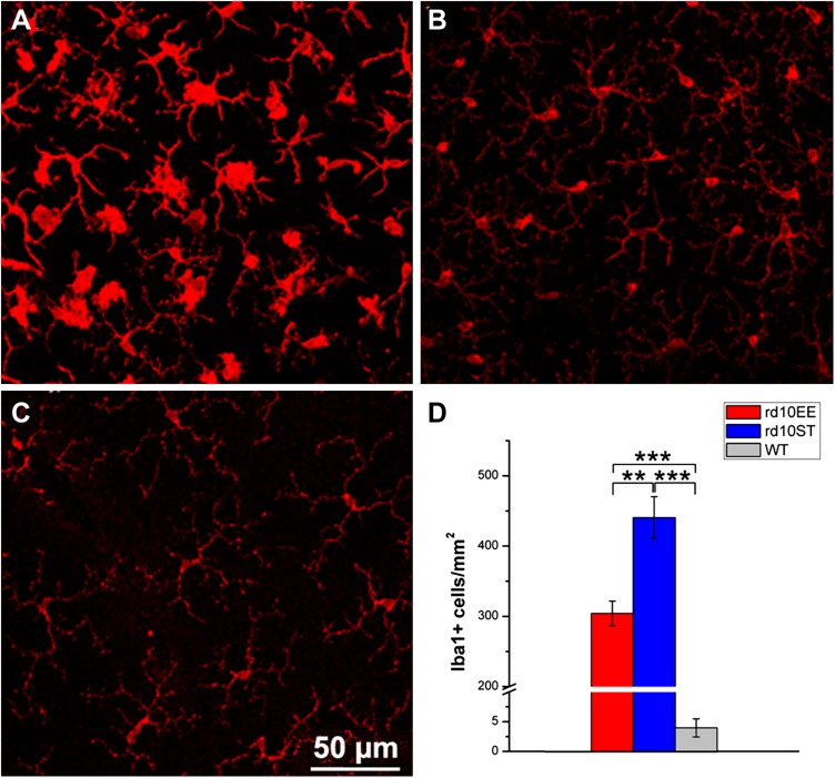
Effects of EE on retinal microglia. A–C) Immunostaining of retinal microglia with Iba1 antibodies at P45. Whole-mount visualization of the outer microglial plexus. rd10, ST retina. Microglial/macrophagic cells have large, ovoid cells bodies and display high reactivity to the immunostaining (A). Processes are short and transition to active status evident. The same preparation from rd10, EE retina shows a high number of Iba1-positive cells with higher ramified morphology and lower activation status (B). In the WT retina, microglial cells and macrophages retain a quiescent morphology and are less numerous than in the diseased retina (C). D) Histograms of Iba-1 positive cells show lower number of microglial cells in EE rd10 retinas compared to ST. The number of these cells is much lower in the healthy, WT retina. **P ≤ 0.01, ***P ≤ 0.001 (ANOVA, followed by Holm-Sidak test).

