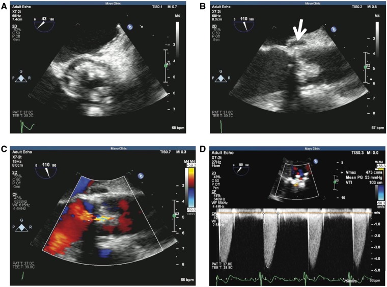
Figure 4.
Transoesophageal echocardiogram of the bioprosthetic valve. (A) Bulky thickening of the prosthetic aortic valve leaflets in short axis. (B) Aortic valve in long axis view showing bulky thickening and echolucency of the aortic root concerning for abscess (arrow). (C and D) Colour flow assessment and Doppler profile of aortic valve prosthesis showing severe stenosis.

