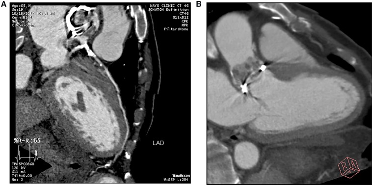
Figure 6.
Cardiac CT angiogram. (A) Severe atherosclerotic disease involving the distal left main coronary artery and proximal LAD with extensive calcification. The distal LAD does not have adequate targets for bypass. (B) Extensive vegetations, primarily on the aortic side of the bioprosthetic aortic valve, with no definite perivalvular extension. PET images (not shown) showed a focal area of increased uptake concerning for an abscess posterior to the valve.

