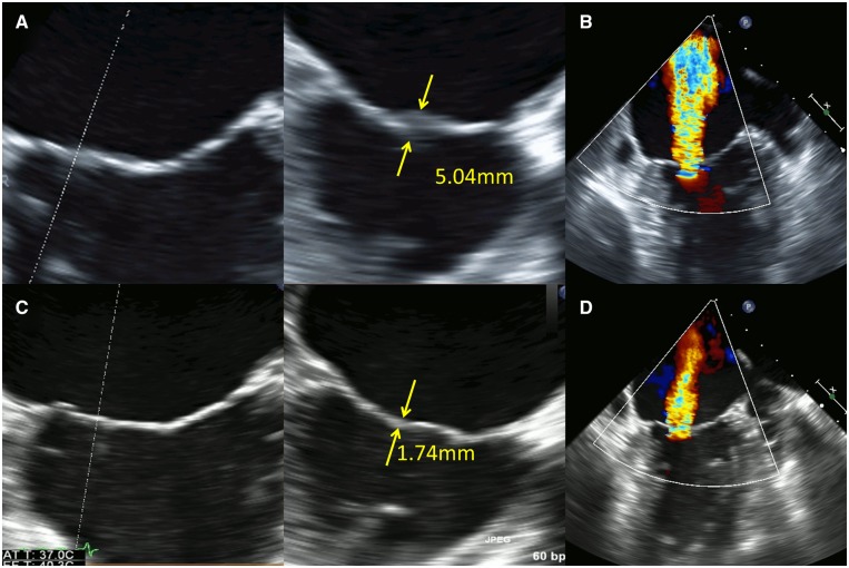
Figure 2.
Transoesophageal echocardiographic images at 14 days (pre-immunosuppressive therapy) showed local low-echoic changes (thickness: 5.04 mm) of the medial edge of A2/P2 segments and the lateral edge of A3/P3 segments with poor coaptation of the mitral valve leaflets, causing severe mitral regurgitation (A and B). Transoesophageal echocardiographic images at 87 days (post-immunosuppressive therapy) elucidated the resolution of the oedematous changes in A2/P2 and A3/P3 segments and thinning of mitral valve leaflets (thickness: 1.74 mm) (C). Mitral regurgitation decreased from severe to mild (D).

