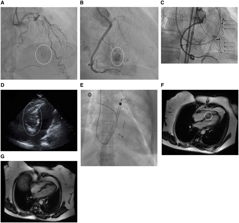
Figure 1.
Fluoroscopic images of the septal haematoma. (A) Initial septal perforation and haematoma formation (circle shows the perforation). (B) Haematoma expansion in the intraventricular septum (circle shows the enlarging haematoma). (C) Coiling of the inflow and outflow of the perforated septal collateral (arrows show the coils). (D) Transthoracic apical four chamber view of the septal haematoma compressing the right ventricle and obliterating the right ventricular cavity (circle shows the haematoma). (E) Fluoroscopic image of an Impella RP device in place with a Swan-Ganz catheter. (F) Baseline cardiac magnetic resonance imaging at discharge showing the septal haematoma (circle shows the haematoma). (G) Repeat magnetic resonance imaging 3 months later with resolution of the septal haematoma.

