
Figure 4.
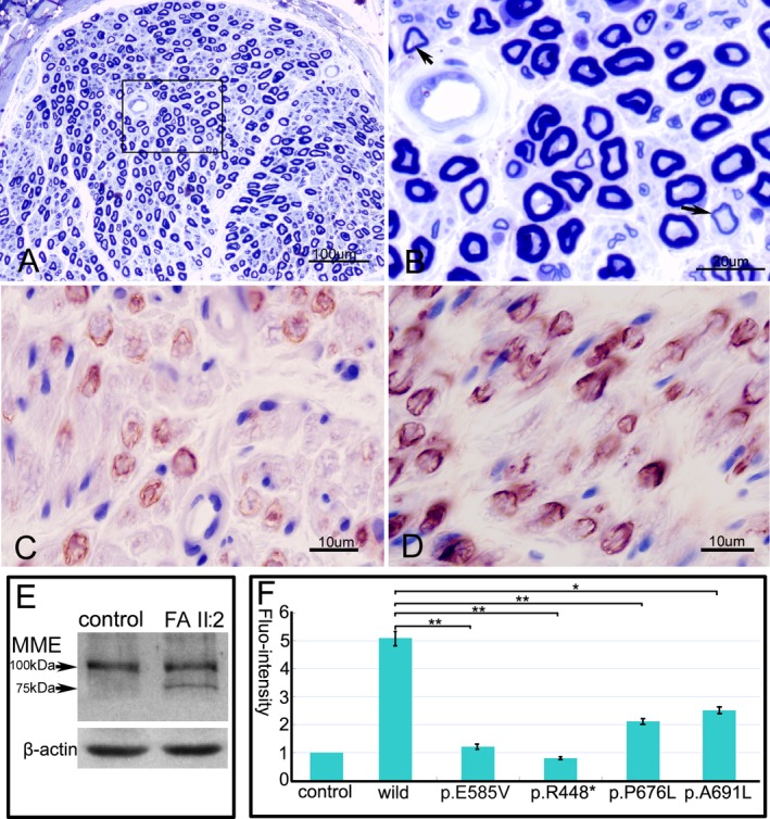
Histopathological findings and expression of MME. (A) Semithin section of sural nerve from the patient II:2 of family A shows a relatively normal density and structure of nerve fibers. (B) The magnification of square in the fig. A reveals few fibers with thin myelin sheaths (arrows). Mild decrease in positive reaction to the nerve fibers in the patient (C) compared to a control (D) by anti‐MME monoclonal antibody. (E) Immunoblot reveals a normal band and a truncated band in the homogenates of sural nerve with c.1342C>T premature mutant. (F) The disease‐associated MME mutants have significant decreases of MME enzymatic activity compared to the wild‐type (*P < 0.05, **P < 0.001). Bars show the mean levels ± standard deviation relative to the empty vector which has been set to +1. MME, membrane metalloendopeptidase.