Figure 6.

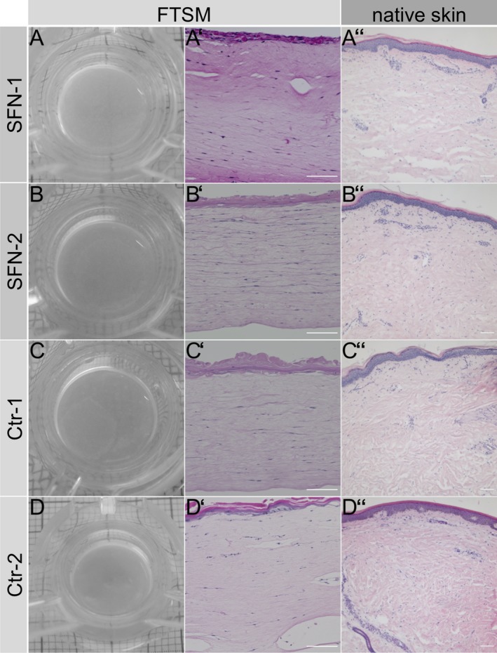
Morphological structure of the full‐thickness skin model (FTSM). Macroscopic images of the FTSM of two patients with small fiber neuropathy (SFN, A, B) and two healthy controls (Ctr, C, D). Hematoxylin and eosin (HE) stainings of the FTSM (A′‐D′), the respective skin punch biopsy (A″–D″) of the patients with SFN, and the healthy control. Morphology was normal in the FTSM of the SFN patients and the healthy controls, and was similar to that of native skin. Scale bar represents 100 μm.
