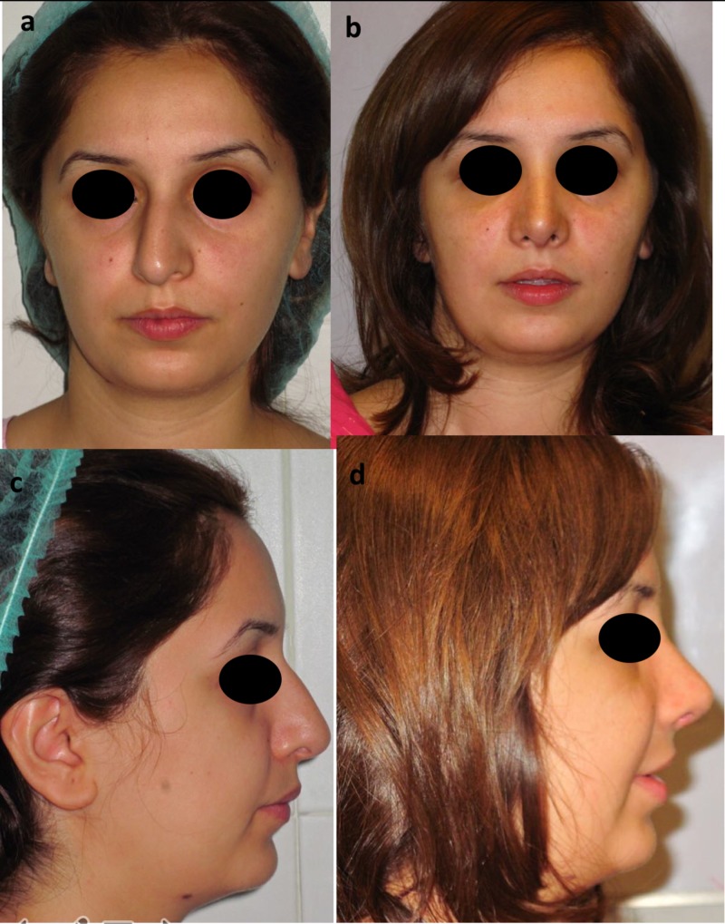
Figure 1. Frontal and lateral views.
Frontal view photographs are shown before (above, left a) and six months after rhinoplasty, dorsal hump reduction, correction of septal deviation, columellar strut, lateral crural strut, interdomal suturing, and spreader graft placement (above, right b).
Lateral view photographs before (below, left c) and after (below, right d) surgery demonstrate a smooth dorsum, good tip elevation, and appropriate nasolabial angle.

