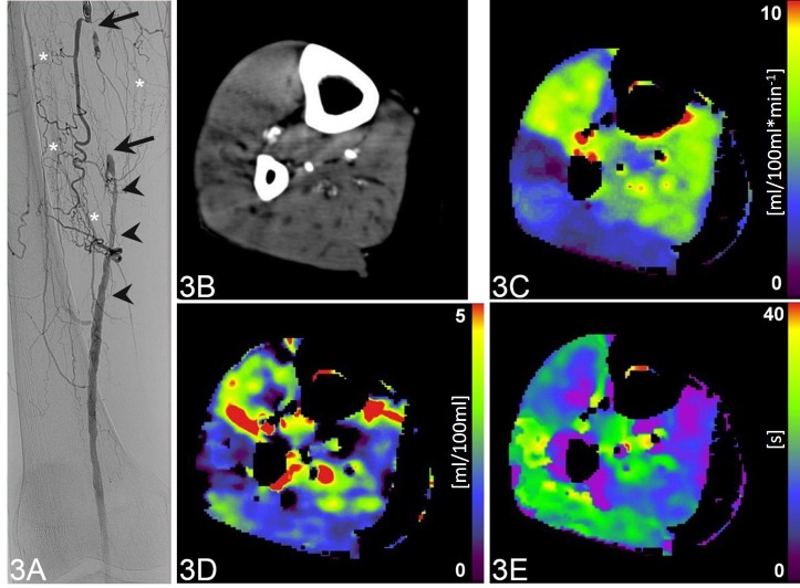
Fig 3. Occlusion of the right-sided superficial femoral artery of a 72-year-old patient.
Coronal digital subtraction angiography image (A) shows a complete intermediate-length occlusion (arrows) of the right right-sided superficial femoral artery, which is extensively collateralized (asterisks). The distal segment of the vessel is patent, however, with caliber irregularities (arrowheads). CTP of the lower leg (B, maximum intensity projection CT image during 60s after contrast administration) with color-coded parametric maps (C-E) reveals a mean blood flow of 5.54 ml/100ml*min-1 (C), mean blood volume of 0.81 ml/100ml (D) and mean transit time of 12.85 s (E).

