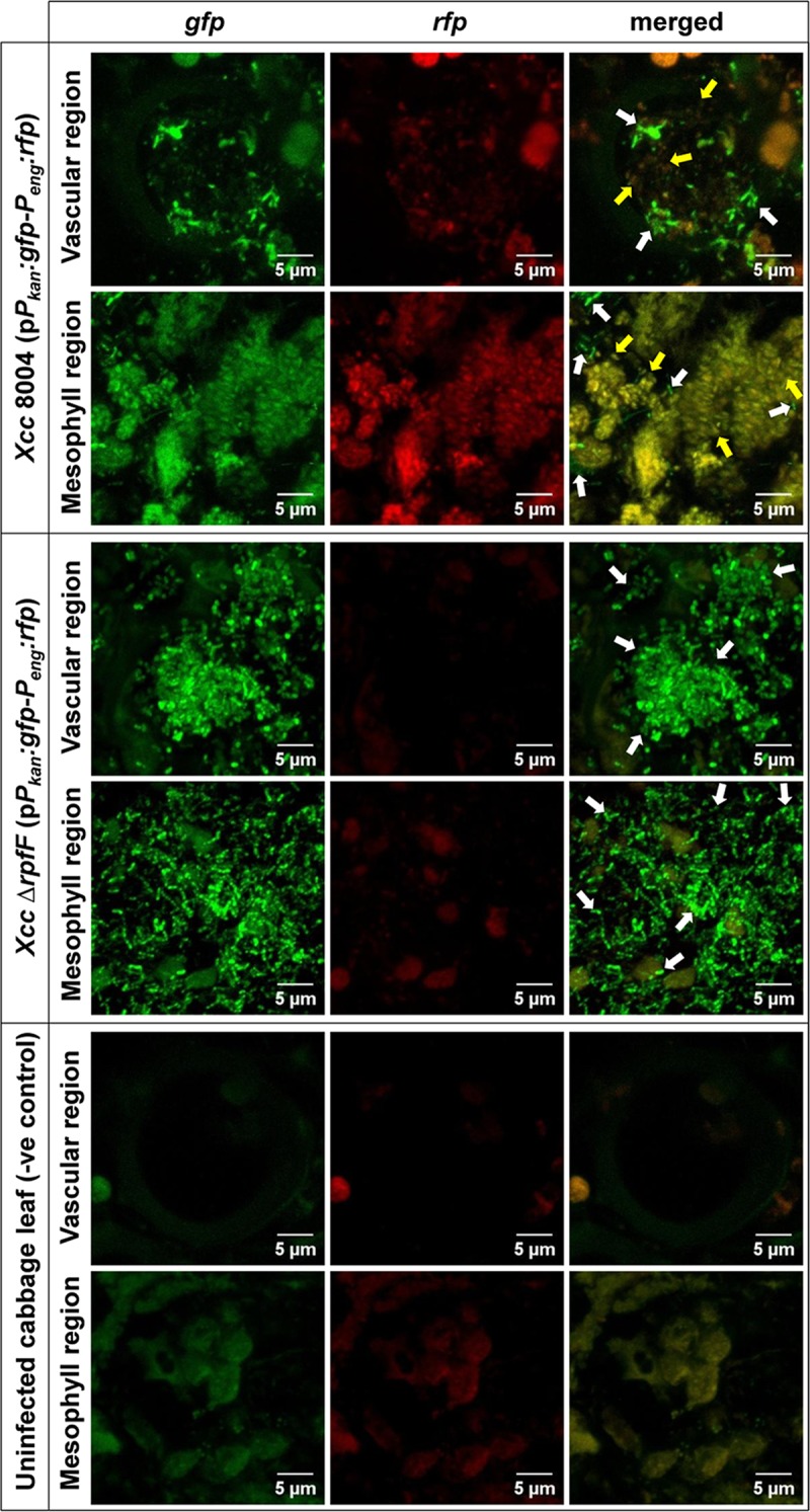
Fig 1. Xcc experiences DSF responsive QS heterogeneity during early stage of disease establishment within host plant.

40 days old healthy cabbage leaves were clipped with low cell density cultures (~ 106 cells ml-1) of the wild-type Xcc-biosensor strain and the inoculated leaves were scanned under CLSM for the QS-induced rfp expression patterns for all constitutive gfp expressing dual-bioreporter cells spanning its proximal non-symptomatic green vascular and mesophyll regions (within 1 cm distance from beyond the diseased symptom periphery) at regular intervals upto 12 dpi. In parallel, its DSF synthesis mutant ΔrpfF-biosensor strain was used as a QS negative control strain. The uninfected cabbage leaves were used as control plant to visualise plant autofluorescence under CLSM at same exposure. Shown in the above figure are the representative CLSM pictures depicting the heterogeneous QS-response within wild-type Xcc 8004 dual-bioreporter populations spanning both vascular and mesophyll regions of infected cabbage leaves on 6 dpi, along with Xcc ΔrpfF (as QS negative control) and uninfected cabbage leaves (as a control plant). The panels from left to right show gfp, rfp and their merged images respectively. For each bioreporter strain, top and bottom panels represent the bacterial populations localizing the transverse sections of individual xylem vessels and mesophyll regions respectively. Yellow arrows; QS-induced cells, White arrows; QS uninduced cells. For uninfected cabbage leaves (i.e. control plant), top and bottom panels represent the plant autofluorescence (without any bacterial populations) for transverse sections of individual xylem vessels and mesophyll regions respectively. Images were prepared using FIJI (image J) software. Scale bars on each panel, 5 μm.
