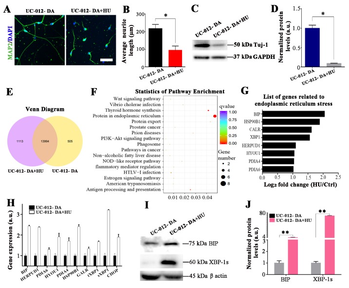
Figure 3.
ER stress was induced by HU treatment in the UC-12-iPSCs-derived dopaminergic neurons. (A, B) The neurite length in the UC-12-iPSCs-derived dopaminergic neurons was dramatically reduced after 4 days of the HU treatment. The neurite was revealed by MAP2 staining. (C, D) Western blot analysis showed that the HU treatment decreased the expression of Tuj1 in UC-12-iPSCs-derived dopaminergic neurons. (E) The number of differentially expressed genes between the control group and the HU treatment group is shown with a Venn diagram. The middle circle indicated the number of mutual expressed genes between the control group and the HU treatment group. (F) The statistics of pathway enrichment analysis for the HU-induced aging in the UC-12-iPSCs-derived dopaminergic neurons. (G) Altered expression levels of genes related to the ER stress pathway. (H) The ER stress related genes were verified by qPCR. (I-J) The expression level of the key proteins in the ER stress pathways was further verified and quantified by western blot analysis. Scale bar: 45 μm for A and 120 μm for C.

