Figure 3.
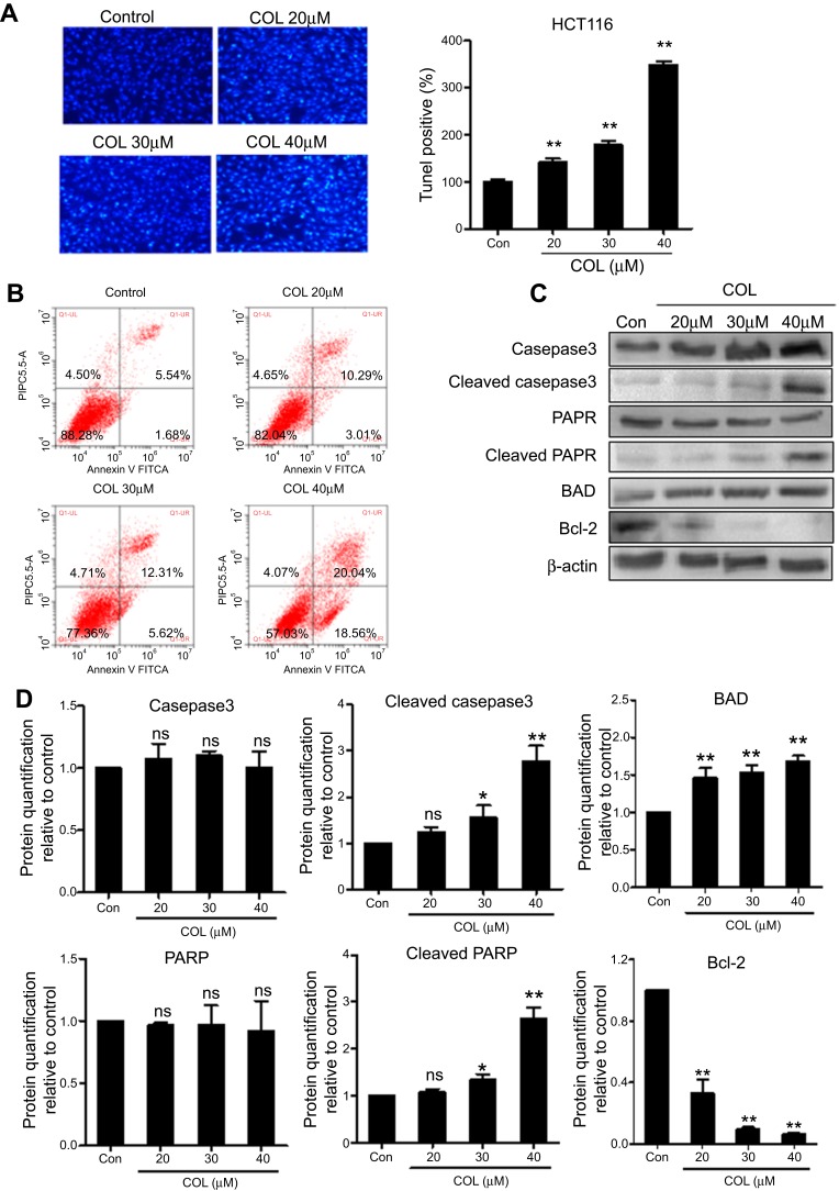
Columbamine induces apoptosis in colon cancer cells. HCT116 cells were incubated with COL at concentrations of 20 μM, 30 μM and 40 μM for 48 hrs. TUNEL staining (A) and flow cytometry (B) were performed to quantify the apoptotic of colon cancer cells. Data were representatives of three independent experiments with similar results. (C) The protein level of apoptosis-related molecules including Caspase3, cleaved Caspase3, PARP, cleaved PARP, BAD, Bcl-2 were detected by Western blotting. (D) Protein levels were quantified by ImageJ software. The gray value of protein band was normalized to its internal control β-actin, and was expressed as values relative to the control from three independent experiments. The error bars represented standard deviations of quantification from three independent experiments.
Notes: ns, P>0.05; **P<0.01; *P<0.05, compared to control group.
Abbreviations: PARP, poly ADP ribose polymerase; BAD, Bcl-2-associated death promoter; Bcl-2,B-cell lymphoma 2; COL, columbamine.

