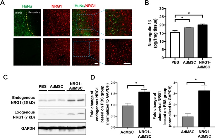
Fig 3. Transplantation of NRG1-expressing AdMSCs increases NRG1 expression in the ipsilateral hemisphere.
(A) Immunohistochemical staining shows that NRG1 immunoreactivity (red) is co-localized with human nuclei (HuNu, green) in the ipsilateral hemisphere whereas no double-labeled cells are detected in the contralateral hemisphere. Scale bar in the low magnified image = 100 μm. Scale bar in the high magnified image = 100 μm. (B) Protein level of NRG1 determined by ELISA is significantly increased by NRG1-AdMSCs transplantation (n = 3 for each group). *p < 0.05 by one-way ANOVA followed by Duncan’s post hoc test. (C) Representative western blot of three independent experiments of exogenous and endogenous NRG1. (D) Graphs showing relative expression levels of endogenous and exogenous NRG1 between AdMSCs- (n = 4) and NRG1-AdMSCs- (n = 6) treated groups. Both endogenous NRG1 (35 kD) and exogenous adenoviral NRG (7 kD) are increased by the transplantation of NRG1-AdMSCs. *p < 0.05 by Student’s unpaired t-test. The data are presented as the mean ± SEM.

