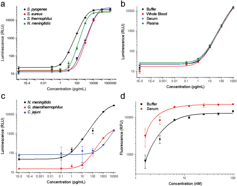
Figure 2. Development of Cas9 protein and activity detection assays.
(a) Detection of Cas9 by capture antibody-functionalized silica microparticles and detection antibodies for S. pyogenes (2 pg/mL), S. aureus (44 pg/mL), S. thermophilus (31 pg/mL), and N. meningitidis (23 pg/mL) using chemiluminescence detection (b) Detection of S. pyogenes Cas9 by capture and detection antibodies have unchanged luminescence profiles regardless of biological matrix. (c) Detection of Cas9 by AcrIIC1-functionalized silica microparticles as a capture reagent. N. meningitidis Cas9 (32 pg/mL) and C. jejuni Cas9 (1293 pg/mL) were detected by species-specific antibodies, while G. stearothermophilus Cas9 (188 pg/mL) could be detected by cross-reactive S. aureus Cas9 antibodies. (d) Microparticle-immobilized fluorescence-based detection of S. pyogenes Cas9 nuclease activity in the presence of the substrate-specific AAVS1 guide RNA. Error bars represent standard deviation, points are average of four replicates, and the limits of detection are indicated in parentheses for each measurement.

