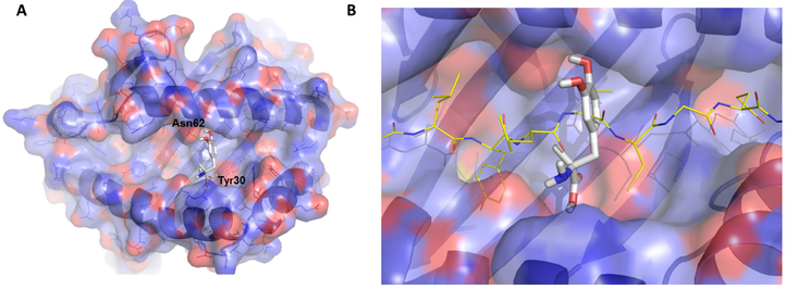
Figure 2.
Molecular models of methyldopa in the DQ8 peptide-binding groove. (A) Methyldopa is shown within pocket 6 of DQ8. There are hydrogen bonds between methyldopa and asparagine (ASN) at position 62 of the DQ8 alpha chain and tyrosine (Tyr) at position 30 of the DQ8 beta chain. These hydrogen bonds were confirmed in structure activity relationship testing using modified methyldopa compounds. (B) Methyldopa in the DQ8 peptide-binding groove clashing with the binding of an insulin peptide. Images were generated using PYMOL software.

