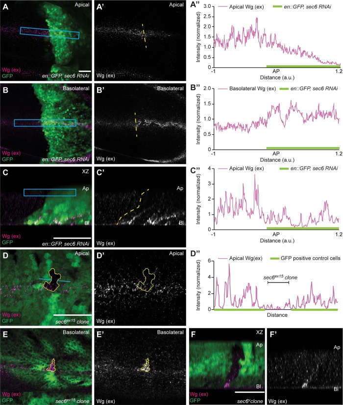
Fig 2. Sec6 depletion blocks apical secretion of Wg.
(A–A”) Representative images of the apical section of extracellular Wg staining performed on disc with Sec6 depletion in the posterior compartment (marked by GFP). Genotype is en-Gal4, UAS-GFP/UAS-sec6-RNAi. (A”) Graph shows normalized intensity profile across the blue box in A. (B–B”) Images of extracellular Wg staining at the basolateral side of the same Sec6 depleted disc. (B”) Graph shows normalized intensity profile across the blue box in B. (C–C”) XZ section of the Sec6 depleted disc shown above. (C”) Normalized intensity profile across the blue box in C. (D–D”) Extracellular Wg staining performed on the discs containing small sec6ex15 mutant clones (marked by absence of GFP) shows modest reduction of apical Wg levels, the graph (D”) shows intensity profile of the extracellular Wg across the blue line shown in D. (E–F) A clear increase in the basolateral Wg can be seen over the same sec6ex15 mutant clone (E), which is also observed in the XZ section (F). N = 3 discs. Scale bar 20 μm, a.u. = arbitrary unit. AP = Anterior-Posterior boundary.

