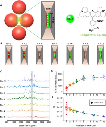
Fig. 5. Quantized single-molecule SERS.

(A) Schematic of the tetrameric metamolecules with accurate number of Raman dye ROX molecules in the hot spot. The diameter of ROX is ~1.6 nm, while the diameter of double-stranded DNA is 2 nm. (B) Schematic of the hot spot region with different numbers of ROX (N = 1, 2, 3, 4, 6, 9, 12). According to the calculated size of hot spot and the diameter of the ROX, six ROX can fill in the hot spot region. (C) SERS spectra taken from seven individual tetramers with different numbers of ROX. (D) Quantized SERS responses as measured by the intensity plot at 1504 cm−1 along with the increase of the number of ROX per particle (N = 12, red, 1 ROX; N = 14, orange, 2 ROX; N = 9, claybank, 3 ROX; N = 9, green, 4 ROX; N = 11, light blue, 6 ROX; N = 8, dark blue, 9 ROX; N = 8, purple, 12 ROX). (E) Measured EFs at 1504 cm−1. All measurements for EF calculations were performed with a 633-nm excitation laser (10-s exposure).
