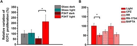
Fig. 2. Polymer-mediated optical activation of TRPV1 stimulates proliferation in ECFCs.

(A) Relative variation of the proliferation rate of ECFCs subjected to long-term optical excitation seeded on both bare glass and P3HT thin films, together with corresponding control samples kept in dark conditions. Cell proliferation was measured after 36 hours of culture in the presence of EBM-2 supplemented with 2% fetal calf serum. (B) Relative variation of the proliferation rate of ECFCs subjected to long-term optical excitation seeded on P3HT in the absence (CTRL) and presence of 10 μM capsazepine (CPZ), 10 μM ruthenium red (RR), 20 μM RN-1734 (RN-1734), and 30 μM BAPTA-AM (BAPTA). The results are represented as the means ± standard error of the mean (SEM) of three different experiments conducted on cells harvested from three different donors. The significance of differences was evaluated with one-way analysis of variance (ANOVA) coupled with Tukey (A) or Dunnett’s (B) post hoc test. *P < 0.05.
