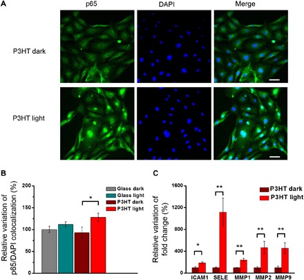
Fig. 6. Light-induced photostimulation promotes p65 NF-κB nuclear translocation and induces the expression of proangiogenic genes in ECFCs.

ECFCs seeded on P3HT samples and glass controls are subjected to long-term photostimulation protocol. Corresponding control samples are kept in dark conditions. After photostimulation, p65 NF-κB nuclear translocation (A and B) and mRNA levels of tubulogenic/angiogenic genes that have been shown to be activated downstream of NF-κB (C) are evaluated. (A) Representative images of immunofluorescence staining showing p65 NF-κB (green) nuclear translocation. Cell nuclei are detected by 4′,6-diamidino-2-phenylindole (DAPI) (blue). Scale bars, 50 μm. (B) Quantitative evaluation of p65 NF-κB nuclear translocation, as evidenced by colocalization experiments. Results are expressed as means ± SEM of the relative percentage of p65 nuclei–positively stained cells to the total number of cells (glass dark, n = 151; glass light, n = 125; P3HT dark, n = 147; and P3HT light, n = 159). Ten fields per condition are analyzed. Data are obtained from two different experiments conducted on cells harvested from two different donors. (C) mRNA levels of intercellular adhesion molecule 1 (ICAM1), selectin E (SELE), and matrix metalloproteinases (MMP1, MMP2, and MMP9) are quantified by real-time polymerase chain reaction (PCR). Data are expressed as means ± SEM of percentage variation with respect to cells grown in the dark (n = 6). The significance of differences was evaluated with unpaired Student’s t test (C) or one-way ANOVA coupled with Tukey post hoc test (B). *P < 0.05 and **P < 0.01.
