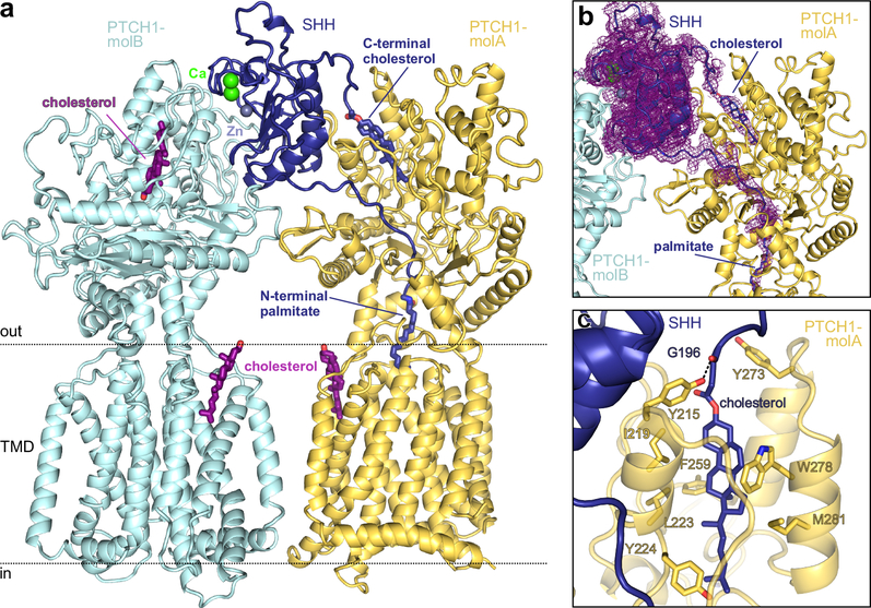
Figure 2 |. Structure of the PTCH1-pShhNc complex.
a, Rebuilt and improved model of the complex between one molecule of pShhNc and two molecules of PTCH1 (molA and molB). The high-affinity protein-protein interface between PTCH1-molB (cyan) and pShhNc is organized by the highly conserved calcium (green)- and zinc (grey)-binding sites of pShhNc (dark blue). The interface with PTCH1-molB (yellow) is composed of the cholesteryl- and palmitoyl- appendages of pShhNc that simultaneously penetrate the ECD. b, The previously deposited cryo-EM map of the SHH-PTCH1 complex (EMD-895516) mapped onto our improved model. Extra density is clearly discernible stretching from the C-terminus of pShhNc into ECD1 of PTCH1. c, Close-up view of the SHH-cholesterol molecule bound in the sterol-binding domain (SBD) of PTCH1, with residues in contact with cholesterol depicted in stick representation.

