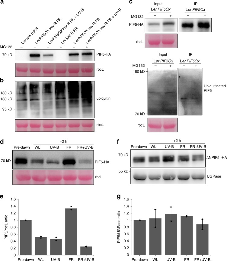
Fig. 4.
UV-B-mediated PIF5 degradation occurs via the proteasome system and requires the APB domain of PIF5. a MG132 inhibits PIF5 protein degradation in LerPIF5Ox. Plants were grown for 10 days in 16 h light/8 h dark cycles before being transferred to ½ strength MS liquid medium containing 0.1% DMSO ± 50 uM MG132 for 16 h. Plants were transferred at dawn to low R:FR ± UV-B for 40 min. PIF5-HA was detected with an anti-HA antibody. Ler was used as negative control and ponceau staining of the Rubisco large subunit (rbcL) was used as loading control. b Western blot of protein samples from (a) probed with an anti-ubiquitin antibody. c Co-IP assay showing PIF5 ubiquitination. Seedlings were grown as in (a). Total protein extracts were immunoprecipitated from low FR + UV-B 30 min treated samples with anti-HA beads and immunoblots probed with anti-HA or anti-Ubiquitin antibodies. Ponceau stained Rubisco large subunit (rbcL) was used as a loading control. d Western blot of PIF5 protein abundance in 35S:PIF5-HA plants. Seedlings were grown for 10 days in16 h light/8 h dark cycles before transfer at dawn to high R:FR ± UV-B for 2 h. PIF5 was detected with an anti-HA antibody. UGPase was used as loading control. e Quantification of PIF5/rbcL ratio in two biological repeats of (d). Bars represent s.e.m. f Western blot of PIF5 protein abundance in 35S:ΔN.PIF5 lines containing a deletion of the first 68 amino acids of the PIF5 protein. Blots were performed as in (c). g Quantification of ΔN.PIF5/UGPase ratio in two biological repeats of (f). Bars represent s.e.m. Source data are provided as a source data file

