Figure 6.
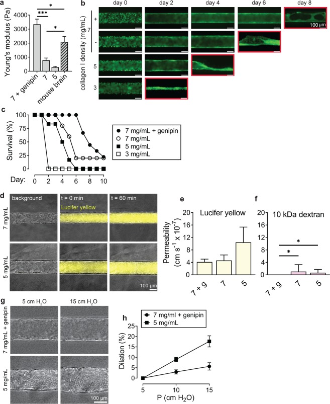
Role of ECM stiffness on structure and function of blood-brain barrier microvessels. (a) Comparison of Young’s modulus for bulk hydrogels: (1) 7 mg mL−1 collagen cross-linked with genipin, (2) 7 mg mL−1 collagen I, and (3) 5 mg mL−1 collagen I, and mouse brain. Data represent mean ± SEM for at least 8 bulk gels across 3 independent preparations, and 5 independent mouse samples. (b) Representative time course images of microvessels for each matrix condition. Monolayers on the softest gel condition, 3 mg mL−1 type I collagen, failed to reach confluence by day 2; such vessels were subsequently failed by collapse of the lumen, as indicated by the red outline on the corresponding time course image. (c) Survival (%) of microvessels over time based on characterization of confluent endothelium and continuous perfusion. Data represents longevity for at least five microvessels across at least 3 independent differentiations. (d) Representative phase/fluorescence overlays of Lucifer yellow permeability of microvessels in 7 mg mL−1 and 5 mg mL−1 collagen I on day 2 following seeding. (e,f) The influence of matrix stiffness on permeability of Lucifer yellow and 10 kDa dextran permeability on day 2. Data represent mean ± SEM for 3 microvessels from at least 2 independent differentiations. (g,h) Dilation of microvessels in response to changes in transmural pressure in 7 mg mL−1 microvessels cross-linked with genipin and 5 mg mL−1 microvessels. Under baseline conditions, the head was 5 cm water corresponding to a transmural pressure of approximately 0.05 Pa. Data represent mean ± SEM for 3 microvessels from 2 independent differentiations.

