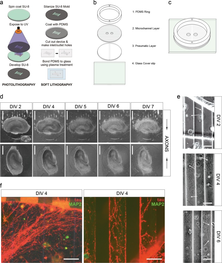
Fig. 2. Microfluidic culture device for high-throughput screening of pharmacological agents to facilitate development of DAI therapeutics.
a Diagram of microfabrication process. b Schematic of the four layer components that comprise the microfluidic culture device. c Schematic of assembled microfluidic device. d Representative phase-contrast images of organotypic hippocampal slice cultures grown in the microfluidics device. Each individual device hosts two organotypic slice cultures, and each slice faces the interconnecting microchannels with the dentate gyrus or CA3 region of the hippocampus, respectively. e Phase-contrast images of axonal projections growing through the interconnecting microchannels between the two culture compartments at various days in vitro. f Fluorescence micrographs of immunostaining against tau protein and MAP2. Processes entering and inside of microchannels are immunopositive for tau and negative for microtubule-associated protein 2 (MAP2). Scale bars, 500 μm (d), 50 μm (e), 50 μm (f)

