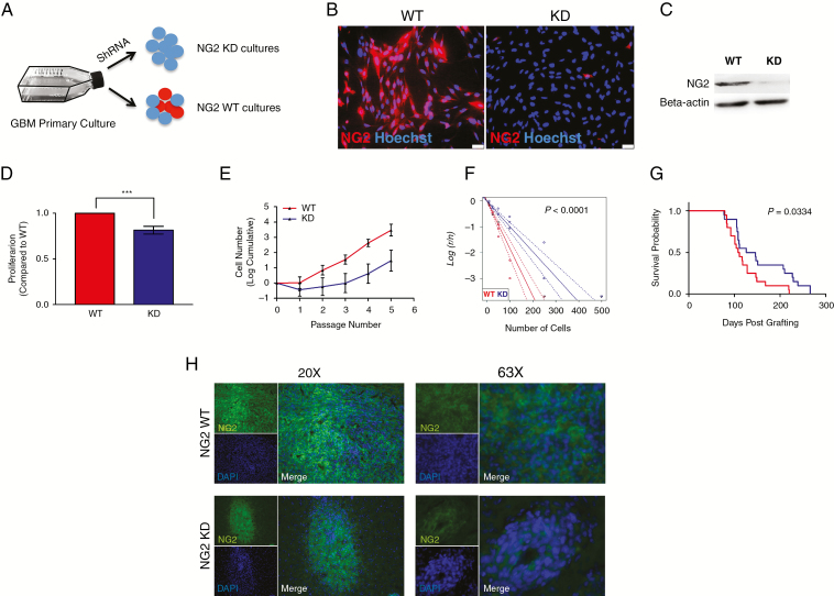
Fig. 3.
NG2 knockdown demonstrates a mechanistic role of NG2 in enhancing GBM cell proliferation. (A) Primary cell cultures were derived from clinical samples before stable NG2 shRNA knockdown. (B) Immunofluorescence staining of NG2 in wild-type (WT) culture and its corresponding NG2 knockdown (KD) culture. (C) Western blot of NG2 from WT and KD cells. (D) BrdU assay conducted on NG2-WT and NG2-KD cells, showing the proportion of proliferating cells. (E) Growth curves over several passages of NG2-WT and NG2-KD cells. (F) Limiting dilution assay (LDA) conducted on NG2-WT and NG2-KD cells. (G) Kaplan–Meier curve showing the survival of experimental animals grafted with NG2-WT or NG2-KD cells. (H) Immunofluorescence staining of NG2 in tumors derived from the injection of WT (upper panels) or KD cells (lower panels). The images show magnification at 20x (left) and 63x (right).

